
As skeletal muscle under microscope 400x takes center stage, this opening passage beckons readers into a world crafted with meticulous precision, ensuring a reading experience that is both absorbing and distinctly original. Delving into the intricacies of muscle tissue, this exploration unveils the fundamental components that orchestrate movement and power, offering a profound understanding of the human body’s remarkable capabilities.
Through the lens of a microscope, we embark on a journey into the realm of skeletal muscle, deciphering its intricate architecture and unraveling the secrets behind its remarkable ability to contract and generate force. Prepare to be captivated as we delve into the fascinating world of muscle histology, where knowledge illuminates the path to a deeper appreciation of human physiology.
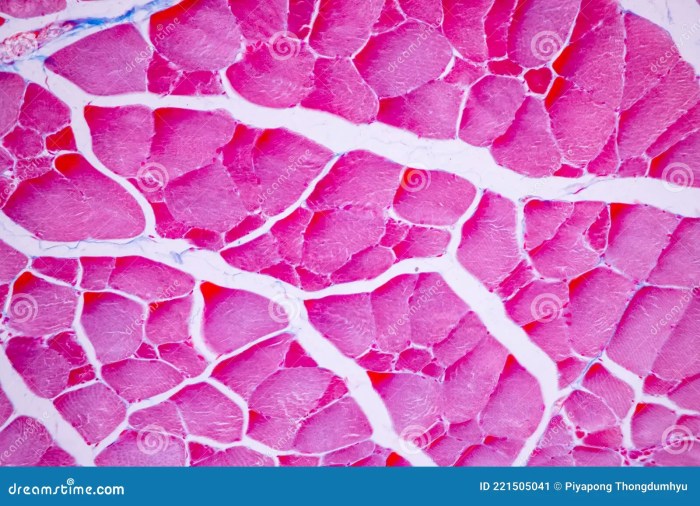
Overview of Skeletal Muscle Histology

Skeletal muscle tissue is composed of elongated, cylindrical cells called muscle fibers. Under a 400x microscope, these fibers exhibit a characteristic striated appearance due to the arrangement of contractile proteins within them. There are three main types of muscle fibers: Type I (slow-twitch, oxidative), Type IIa (fast-twitch, oxidative-glycolytic), and Type IIx (fast-twitch, glycolytic).
Myofibrils and Sarcomeres
Within muscle fibers, contractile proteins are organized into myofibrils, which are long, thin structures that run parallel to the long axis of the fiber. Myofibrils are composed of repeating units called sarcomeres, which are the basic units of muscle contraction.
Sarcomere Structure
Each sarcomere is bounded by two Z-lines, which are composed of the protein alpha-actinin. The central region of the sarcomere, called the A-band, contains thick myosin filaments. The I-band, located on either side of the A-band, contains thin actin filaments.
The H-zone is the central region of the A-band where only thick filaments are present, while the M-line is a thin line in the middle of the H-zone where thick filaments are attached.
Myofilaments and Cross-Bridges
Myofilament Composition
Thick myosin filaments are composed of myosin molecules, which have a head and tail region. The head region contains ATPase activity and can bind to actin filaments. Thin actin filaments are composed of actin molecules, tropomyosin, and troponin. Tropomyosin molecules block the binding sites on actin for myosin heads, while troponin molecules regulate the interaction between actin and myosin.
Cross-Bridges
During muscle contraction, myosin heads extend and bind to actin filaments, forming cross-bridges. The formation of cross-bridges allows the thick and thin filaments to slide past each other, shortening the sarcomere and generating force.
Sarcoplasmic Reticulum and T-Tubules

Sarcoplasmic Reticulum
The sarcoplasmic reticulum (SR) is a network of tubules that runs throughout the muscle fiber. It stores calcium ions, which are released into the cytosol to trigger muscle contraction. The SR is divided into two types: terminal cisternae, which are located near the T-tubules, and longitudinal tubules, which run parallel to the myofibrils.
T-Tubules
T-tubules are invaginations of the sarcolemma (cell membrane) that run perpendicular to the myofibrils. They allow the action potential from the sarcolemma to spread rapidly throughout the muscle fiber, triggering the release of calcium ions from the SR.
Nerve-Muscle Junction: Skeletal Muscle Under Microscope 400x
Structure
The neuromuscular junction (NMJ) is the site where a motor neuron connects to a muscle fiber. The motor neuron releases the neurotransmitter acetylcholine (ACh), which binds to receptors on the muscle fiber’s sarcolemma. This binding triggers the opening of ion channels, leading to an action potential that spreads throughout the muscle fiber.
Muscle Contraction, Skeletal muscle under microscope 400x
The action potential triggers the release of calcium ions from the SR, which bind to troponin molecules on actin filaments. This causes a conformational change in troponin, which uncovers the binding sites on actin for myosin heads. Myosin heads can then bind to actin and initiate muscle contraction.
Muscle Capillaries and Blood Supply
Capillary Distribution
Skeletal muscle tissue is highly vascularized, with a dense network of capillaries surrounding each muscle fiber. Capillaries are the smallest blood vessels and allow for the exchange of nutrients, oxygen, and waste products between the blood and muscle fibers.
Blood Flow
Blood flow to skeletal muscle is regulated by a variety of factors, including neural input, metabolic demand, and hormones. During exercise, blood flow to skeletal muscle increases significantly to meet the increased metabolic demands.
Commonly Asked Questions
What is the significance of striations in skeletal muscle?
The striated appearance of skeletal muscle, visible under a microscope, arises from the precise arrangement of myofibrils and sarcomeres, which are the fundamental units of muscle contraction.
How do nerve impulses trigger muscle contraction?
Nerve impulses travel along motor neurons and release neurotransmitters at the neuromuscular junction, which triggers an action potential in the muscle fiber. This action potential initiates the release of calcium ions from the sarcoplasmic reticulum, leading to muscle contraction.
What is the role of blood capillaries in skeletal muscle?
Blood capillaries provide oxygen and nutrients to muscle fibers and remove waste products. They also play a crucial role in regulating muscle temperature and pH levels.
